

Capsule Endoscopy
Since its introduction into clinical use in 2001, capsule endoscopy has been a game-changer by allowing gastroenterologists to non-invasively image the entire small bowel. This technology has not only earned clinical trust but is constantly being refined by integrating artificial intelligence into this revolutionary technology.
The journey of capsule endoscopy from fantasy to clinical reality began in 1981, when Gavriel Iddan, a military engineer, and Eitan Scapa, a gastroenterologist, addressed the challenges of examining the small intestine using fiber-optic methods. Their work and that of many talented physicists and engineers eventually merged with the efforts of Paul Swain, who independently experimented with the wireless transmission of mini-cameras.
Clinical use began in 2001, and the first international conference on capsule endoscopy was held in 2002. The clinical success, tolerability, convenience, and safety of capsule endoscopy have inspired the investigation of video capsules beyond the small bowel and into the esophagus, stomach, and colon.
What Is a Capsule Endoscopy?
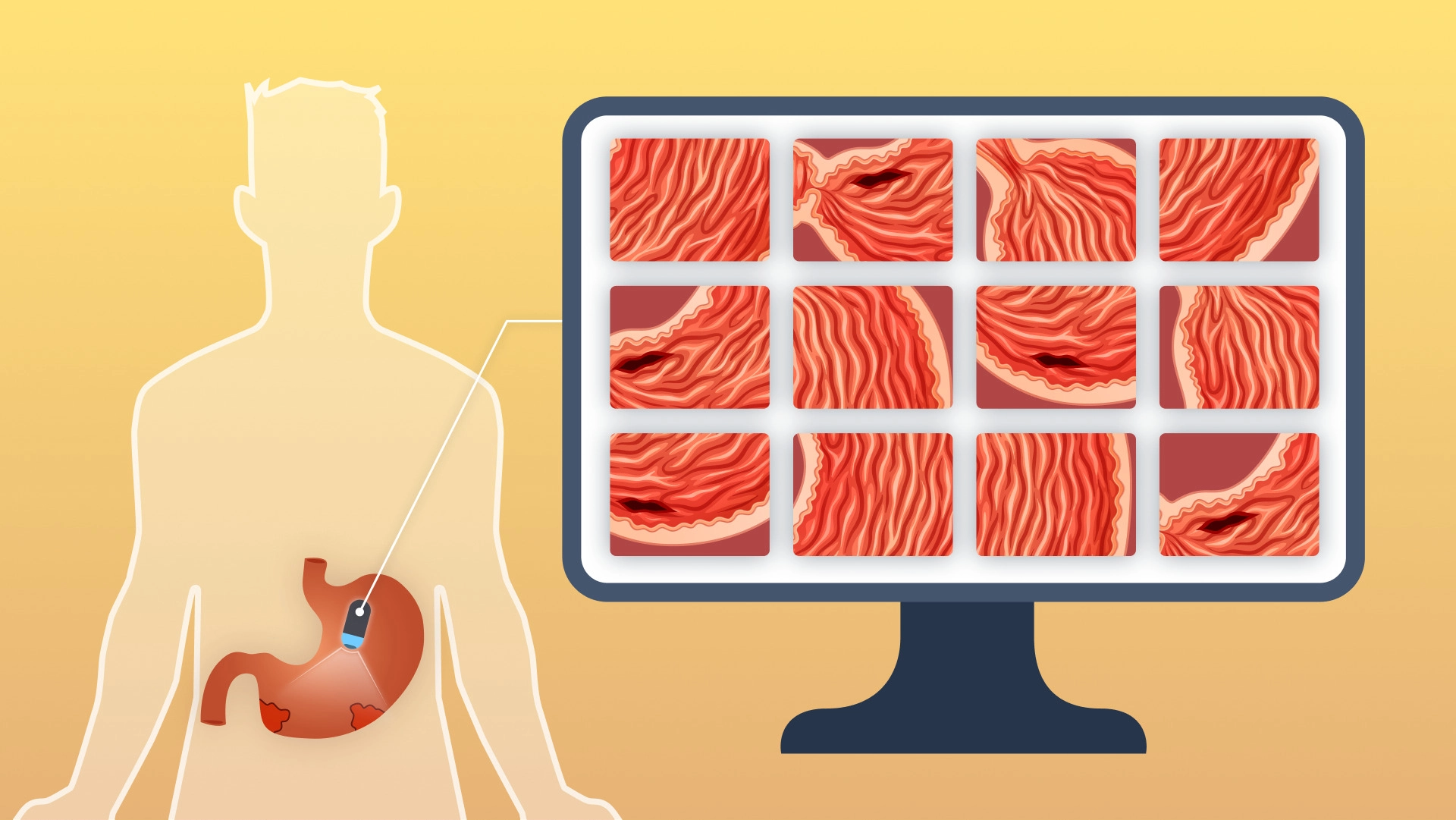
Capsule endoscopy (CE) is a perfect alternative to classical endoscopy: it’s a non-invasive diagnostic procedure for visualizing the interior of the digestive tract. The patient swallows a capsule that contains a tiny camera, a transmitter, and a light. Passing through the stomach, intestines, colon, and rectum, the capsule takes thousands of pictures and transmits them to a recorder that a person wears outside the body.
What Conditions Can Be Diagnosed Using Capsule Endoscopy?
Capsule endoscopy can detect signs of digestive problems and help diagnose a variety of conditions. A doctor may recommend a capsule endoscopy if it is suspected that a person has:
Crohn’s disease and other forms of inflammatory bowel disease (IBD);
gastrointestinal bleeding;
celiac disease;
ulcerative colitis;
colon polyps or colon and rectal cancer.
CE can help the doctor:
confirm a suspicion of Crohn’s disease;
monitoring of celiac disease;
investigate causes of unexplained weight loss or anemia.
Capsule endoscopy can also help doctors visualize the inside of the colon in people who cannot tolerate sedation or other aspects of colonoscopy.
Evolution of Technology
The capsule endoscope has evolved over the years. The first-generation PillCam SB1 capsule (Given Imaging) had a field of view of 140 degrees, a battery life of 8 hours, and a shooting speed of 2 frames per second (fps). PillCam SB2 increased the field of view to 156 degrees and improved optics. The newest version of the PillCam SB3 (now Medtronic) includes adaptive frame rate technology (up to 6 frames per second) that can adjust the image capture speed depending on how fast the capsule is moving.
Other developers have introduced versions of capsules with unique functions. The OMOM (Jinshan) smart capsule has two-way data transmission via a portable RF sensor that allows real-time adjustment of light intensity and capture rate. Compared to the PillCam SB3, it demonstrated similar diagnostic performance with a shorter boot time, but significantly shorter battery life. Other capsules have an increased viewing angle, such as 170 degrees in the MiroCam (IntroMedic), or an increased battery life of up to 12 hours, as in the CapsoCam (Capsovision). The implementation of magnetically assisted upper gastrointestinal capsule endoscopy (Mcapsule Endoscopy) in the small intestine has also been evaluated and found to be well tolerated with good sensitivity to lesions. With the help of an external magnet, the doctor can control the capsule to obtain a focused examination.
Capsule endoscopy does not allow tissue sampling or therapy. However, work is underway to develop capsules that can recognize lesions, fixate and obtain tissue samples. In addition, real-time viewing and maneuverability may allow targeted delivery of drugs or blood-stopping sprays.
Capsovision has developed a new approach to imaging the small intestine with the CapsoCam: a 360-degree capsule. Unlike the standard single-camera approach, this capsule uses 4 cameras, each with a field of view of about 90 degrees. CapsoCam has 15 hours of battery life and a frame rate of 20 frames per second. It also differs from other capsules in that the capsule itself acts as a storage system. No need for a voice recorder, sensor belt, or wires. However, patients should receive the capsule after it has left the body.
Although CE was first introduced 21 years ago, it was not popularized and the number of all producers on the market is still less than 10, with one of them coming from Europe — Biocam. The startup reached up to 97% accuracy of neural networks in detecting images in preclinical trials. Thanks to the algorithms, they are able to increase the image resolution up to 4 times. Additional help for the patients is Biocam’s convenient mobile app which tells all the required instructions about prep for the examination and reminds people about all the steps needed. You can learn more about Biocam’s approach to capsule endoscopy and its startup development in our interview with Biocam’s machine-learning specialist Amelia Okulewicz.
Why Did CE Not Become the Only Technology Used?
Although the capsule provides the best way to view the interior of the small intestine, there are several limitations and problems with its use, the most important of which is that the capsule does not allow for therapy. With the help of a classic endoscope, in addition to its basic functions, certain medications can be introduced into the intestine. Among other disadvantages:
Abnormalities in some areas of the intestine are missed due to the rapid passage of the capsule.
CE is an all-day test, although patients do not usually stay in the hospital to complete it.
If there are narrow areas in the small intestine due to scarring or tumors, the capsule can get stuck in and cause intestinal obstruction, needing surgical removal of the capsule. (For this reason, patients suspected of having this diagnosis first swallow a self-dissolving dummy capsule. If it sticks, this can be seen on an abdominal X-ray and the site of the narrowing can be identified.)
After all, reviewing tens of thousands of photos takes a lot of time for a conscientious doctor. That is why the main attention of developers should be paid to artificial intelligence, which is capable of qualitatively lowering the time required for viewing and analyzing images and improving the expert’s accuracy.
Artificial Intelligence & CE
Reading capsule endoscopy studies can take a long time, and the time ranges from 30 to 120 minutes. A typical capsule may contain 50,000 to 60,000 images from a single study, and abnormalities may be present in only 1-2 frames. In addition, due to the uncontrolled and non-linear movements of the capsule, many images are duplicated, and this redundancy adds time to image review. Software algorithms were developed to account for this redundancy and help reduce read times. A recent study compared rapid reading software and conventional reading for the diagnosis of small bowel lesions. The software algorithm reduced the reading time by 40% (average time savings of 24.6 minutes), without reducing clinical accuracy. Artificial intelligence has also been developed to help identify erosions, ulcers, bleeding, and celiac disease. During studies, a computed detection algorithm showed 100% sensitivity and 96% specificity in assessing state changes.
The Future of Capsule Endoscopy with AI
New artificial intelligence systems that have been developed to assist clinicians and facilitate the interpretation of CE are constantly being developed. An ideal AI system for pathology detection in CE video should have the following characteristics:
• excellent performance (high sensitivity/specificity/accuracy and meager error rate);
• capability to detect multiple lesions;
• ability to classify lesions;
• short video reading time;
• ease of use.
The future of capsule endoscopy is extremely interesting and promising. Technological advances will lead to new, upgraded navigable pods, while AI will provide digital health computing systems that can review CE videos, find possible lesions, and form a diagnosis in less than 30 minutes. When this combination is introduced into daily practice, the impact on endoscopy will be enormous. With the help of just one capsule, it will be possible to accurately and quickly examine the entire gastrointestinal tract. Whether this future is far or near remains to be determined, since the emergence of this new technology depends on the interaction of several factors, including scientific, economic, and managerial aspects.
Final Word
Since its humble beginnings as a noninvasive way to examine the small bowel, capsule endoscopy has become an integral component in the investigation of small bowel disorders. It will continue to improve accuracy and reduce reading times. These innovations may lead to non-invasive biopsy or targeted drug delivery, further expanding the reach and role of capsule endoscopy.

Tell us about your project
Fill out the form or contact us

Tell us about your project
Thank you
Your submission is received and we will contact you soon
Follow us