
Transforming Medical Imaging With Artificial Intelligence
- The Algorithms Behind the Curtain: How AI Actually Works in Imaging
- Real-World Impact: AI That's Already Saving Lives
- New Clinical Workflows and Professional Roles
- From Prototype to Practice: Why Most Imaging AI Fails to Scale—and How to Fix It
- The Next Chapter: Multimodal AI and the Redesign of Diagnostics
Imagine a busy urban hospital where a radiologist is tasked with reviewing over 100 chest CT scans in a single shift. By the time they reach scan number 87, fatigue begins to set in — a common and well-documented issue in diagnostic medicine. In one such case, a subtle pulmonary embolism was nearly missed — until an AI system flagged it in real time. The tool had been integrated into the workflow as a second reader, scanning each image in the background. The alert prompted a second look — and saved a life.
This scenario is not rare or speculative. It's becoming increasingly common in hospitals that are deploying FDA-cleared AI tools like Aidoc to support the detection of critical findings such as pulmonary embolisms, brain hemorrhages, and cervical spine fractures. A 2024 study published in PubMed showed that the implementation of an AI algorithm significantly reduced the rate of missed iPEs from 50% to 7.1%, thereby enhancing diagnostic accuracy.
The growing demand for imaging, combined with a chronic shortage of trained radiologists, has created a pressure cooker environment in diagnostic medicine. According to the Royal College of Radiologists, imaging demand in the UK alone has risen by more than 30% over five years, while workforce growth has lagged behind. Meanwhile, each CT scan can generate hundreds of slices, and large hospitals produce terabytes of imaging data annually — much of it under-analyzed or entirely overlooked due to time constraints.
Artificial intelligence offers not just a way to cope, but a chance to fundamentally redesign how diagnosis happens. AI systems don’t get tired. They don’t miss details due to a long shift or a momentary lapse in focus. And crucially, they can prioritize urgent cases — often before a human even sees them.
In this new reality, AI is not replacing the radiologist — it’s transforming their role and reshaping diagnostic pathways.
The Algorithms Behind the Curtain: How AI Actually Works in Imaging
At the heart of modern AI-powered imaging systems are convolutional neural networks (CNNs), a type of deep learning model designed to analyze visual data by learning hierarchical features from images. In clinical practice, these algorithms are trained to recognize subtle patterns in X-rays, CT, or MRI scans—often patterns that are too small or complex for even experienced radiologists to consistently detect.
CNNs work by passing an image through a series of layers, each extracting different levels of abstraction—from basic shapes and edges in early layers to complex structures like tumors or vascular abnormalities in deeper layers. Unlike traditional machine learning models that require manual feature engineering, CNNs learn directly from raw image data, making them particularly effective in tasks like classification, segmentation, and anomaly detection. For example, U-Net architectures are commonly used to segment lesions in CT scans, while deeper networks like ResNet and EfficientNet power classification models that flag suspected disease areas in chest X-rays.
These systems have shown impressive performance. Many models now match or exceed radiologist-level accuracy in specific diagnostic tasks, with sensitivity and specificity often ranging between 85% and 95%, depending on the condition and dataset. For example, in a separate study published in PLOS ONE in 2021, researchers developed an ensemble of three CNNs (GoogLeNet, ResNet‑18, DenseNet‑121) to detect pneumonia using two public datasets (Kermany and RSNA), achieving 98.81% accuracy and 98.80% sensitivity on the Kermany dataset, with similarly strong performance across metrics on the RSNA dataset. Speed is another advantage—AI can process hundreds of scans in seconds, enabling real-time triage in emergency departments.
Yet, this power comes with critical limitations. One of the most pressing challenges is the so-called “black box” nature of CNNs. These models often do not provide clear reasoning for their predictions, making them difficult to trust in high-stakes clinical settings. This concern has driven the emergence of explainable AI (XAI), a subfield focused on increasing the interpretability of deep learning systems. Tools like Grad-CAM and LayerCAM help visualize which areas of an image influenced the model’s output, allowing clinicians to validate that the algorithm is focusing on medically relevant regions. Others have taken a more integrated approach, embedding attention mechanisms directly into models to enhance interpretability from the outset.
Another concern is generalizability. Models trained on high-quality datasets from a few large hospitals may perform poorly when deployed in different regions, on different scanner types, or with more diverse populations. A study in Diagnostics found that even small differences in image quality or patient demographics can significantly impact model accuracy, particularly for underrepresented populations (MDPI, 2023). This ties into a deeper issue: algorithmic bias. Research published in Nature Medicine and reported by Wired demonstrated that some deep learning systems could infer a patient’s race from X-ray images—even though this information was invisible to human radiologists—which raises serious ethical questions about data leakage and unintended discrimination (Wired, 2022).
Despite these challenges, the core architecture of CNNs continues to underpin nearly every major AI system in imaging today. Their ability to scale, learn from millions of examples, and operate at machine speed makes them indispensable. But as clinical deployment grows, so does the pressure for these models to not only be accurate, but also explainable, robust, and fair across patient populations. In this context, algorithm design is no longer just a matter of performance—it’s a matter of trust.
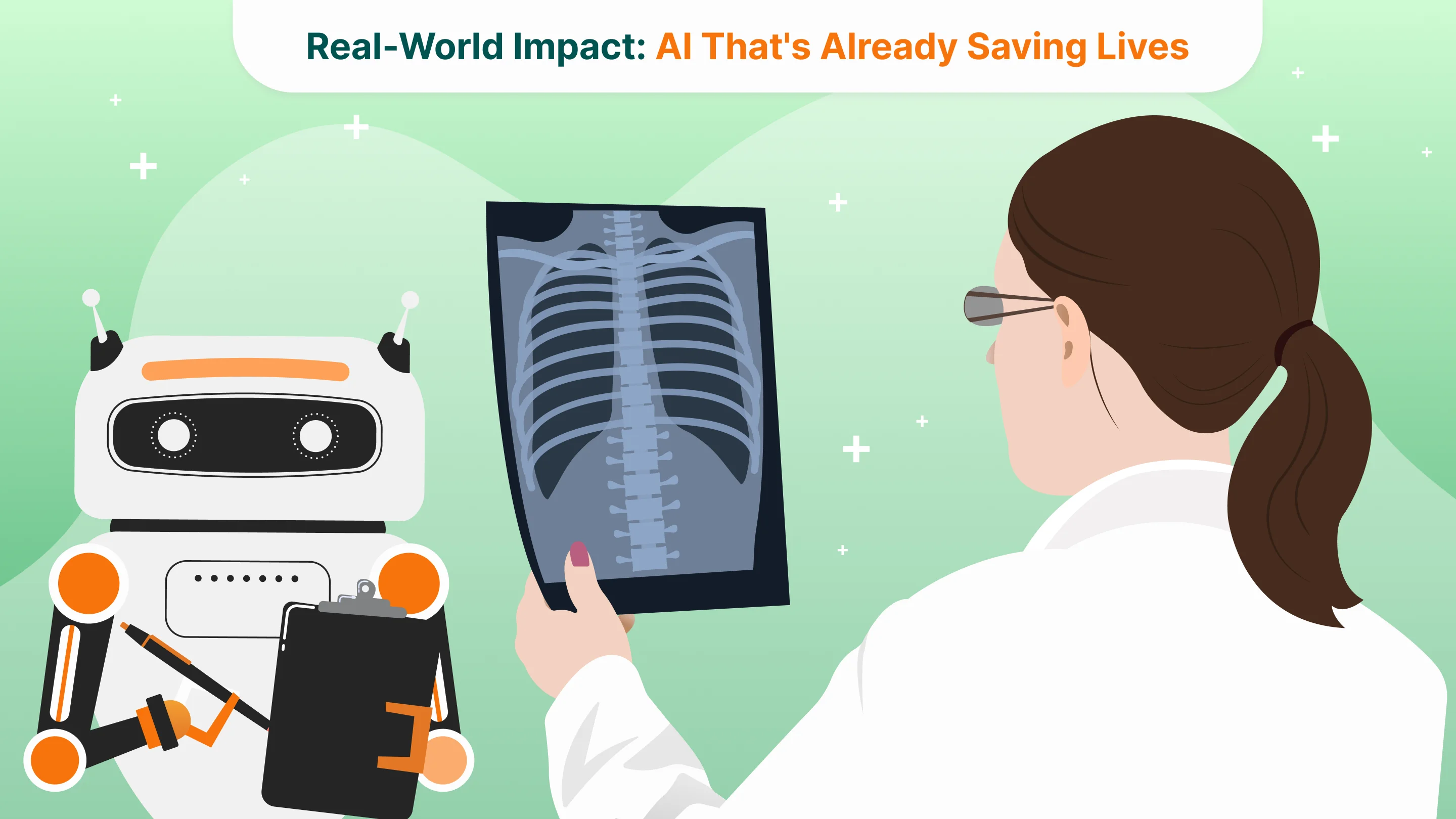
Real-World Impact: AI That's Already Saving Lives

Several AI-driven companies are no longer in the realm of theory—they’re actively reshaping clinical workflows and improving patient outcomes today.
One notable example is Aidoc’s FDA-cleared algorithm for incidental pulmonary embolism (PE). In a study at the Netherlands Cancer Institute involving 11,736 contrast-enhanced chest CT scans, researchers found that, before deploying AI assistance, radiologists missed 44.8% of incidental PEs. After integration with Aidoc, the miss rate dropped dramatically to just 2.6%—a 94% reduction in overlooked cases (Radiology: Cardiothoracic Imaging, 2023/Diagnostic Imaging, 2023) A retrospective single-center observational study also reported that AI assistance improved sensitivity from 50% to 90% in detecting incidental PEs, while maintaining specificity at 99%, significantly reducing radiologist oversight of critical findings (Aidoc clinical study).
Aidoc’s technology isn’t limited to pulmonary embolism; its products are now deployed in over 900 hospitals and have been cleared by both the FDA and CE for detecting intracranial hemorrhages, spinal fractures, free abdominal air, and more. This illustrates how a startup can transition from innovation to infrastructure, integrating AI into Picture Archiving and Communication Systems (PACS) so radiologists receive automated alerts and can reorder priorities in real time.
In another context—public health screening—Qure.ai’s qXR has emerged as a frontline tool. During a Health Technology Assessment by the Indian Institute of Public Health, Gandhinagar, qXR demonstrated superior cost-efficiency over traditional clinical pathways for TB screening. It not only improved diagnostic yield but also reduced per-case costs, proving a truly sustainable solution for low-resource settings. Deployed across India’s rural and urban TB programs—including Mon District, Nagaland—qXR has enabled non-specialist clinicians to detect TB with minimal delay and without onsite radiologists, driving 30–40% increases in TB notifications and 15.8% increases in overall diagnostic yield due to AI alone.
These rapid screening initiatives are supported by rigorous evidence. A retrospective international study across 10 countries, including India and South Africa, benchmarked deep-learning systems against radiologists for TB detection via chest X‑rays. The automated tool achieved an AUC of 0.90 (95% CI: 0.87–0.92), with a sensitivity of 88% vs. radiologists’ 75%—notably reducing screening costs by 40–80% by optimizing confirmatory testing pathways.
New Clinical Workflows and Professional Roles
AI is not simply an add-on—it is actively reshaping diagnostic processes and altering how radiologists work every day. In many modern hospitals, AI tools automatically triage scans as they arrive, flagging urgent findings like intracranial hemorrhage or pulmonary embolism, and allowing clinicians to sequence cases based on severity.
For instance, Aidoc’s solutions are now integrated into PACS systems in over 900 hospitals, delivering real-time alerts that significantly speed up case acquisition and decision-making. In breast cancer screening, Lunit’s AI-CAD has demonstrated operational and diagnostic impact: in South Korea’s single-reader national screening program, AI-assisted radiologists detected 13.8% more cancers without increasing recall rates, and when used as an independent reader in Sweden, it enabled a reduction of one human reader while maintaining or improving detection accuracy (cancer detection rate 5.70 vs. 5.01 per 1,000; recall rate stable at ~6.9%).
These changes shift the radiologist’s role from primary “image reader” to clinical orchestrator—verifying AI-flagged cases, interpreting model explanations, and focusing human expertise where it matters most. At the same time, a growing subspecialty of “AI validation and governance” has emerged: clinicians and data scientists collaborate to tune AI thresholds, evaluate performance across populations, and ensure ongoing safety. In busy screening programs, AI streamlines routine reads, allowing radiologists to dedicate more time to complex cases, consulting with multidisciplinary teams, and directly advising on patient treatment plans. In effect, AI is translating raw image data into structured, actionable insights, enabling radiologists to operate at a higher level—strategic, advisory, and integrative—within the care continuum.
From Prototype to Practice: Why Most Imaging AI Fails to Scale—and How to Fix It
Building a medical imaging algorithm that works in a controlled environment is one thing. Getting it deployed in hundreds of hospitals, used daily by clinicians, and trusted by healthcare systems is something else entirely. Despite the explosion of AI tools in radiology, only a fraction have reached meaningful adoption. The reasons lie not in model accuracy, but in scalability friction.
First, many startups underestimate how complex hospital IT ecosystems are. Integrating into PACS, RIS, and EHR systems often requires deep customization, vendor cooperation, and compliance with regional procurement rules. Even the best-performing model will be sidelined if it can’t run seamlessly within a radiologist’s daily workflow. That’s why companies like Aidoc and Lunit have prioritized system-level compatibility and zero-click integration, enabling alerts to appear directly in the imaging stack, without clinicians opening another tab.
Second, clinical validation is often misunderstood. A solid ROC curve on public data isn’t enough. Hospitals want local evidence: “Does this work on our patients, our scanners, with our doctors?” Leading companies now deploy site-specific tuning and shadow deployments, allowing the model to run silently in the background, comparing its predictions to radiologist reports over weeks or months before going live. This not only builds internal buy-in but also surfaces edge cases and necessary recalibrations.
Another overlooked challenge is reimbursement. In many regions, there is no billing code for AI-assisted diagnosis, meaning hospitals must absorb the cost themselves. Startups that secure CPT codes or demonstrate clear return on investment through time savings, error reduction, or reduced re-scans are more likely to cross the commercial adoption gap.
But perhaps the most underestimated factor is trust-building. Clinicians don’t want another dashboard—they want a partner that helps them make better decisions. That’s why explainability matters not only in theory, but in interface design: visual overlays, real-time confidence scores, and side-by-side comparison tools are no longer nice-to-haves, but core components of adoption. As startups move from academic prototypes to enterprise-scale deployment, the game shifts from "Can this detect cancer?" to "Can this make my workflow easier, safer, and faster—without slowing me down?"
In this landscape, the most successful companies aren’t just great at AI—they’re great at deployment, support, clinical collaboration, and infrastructure thinking. That’s where the next generation of imaging startups will win or lose.
The Next Chapter: Multimodal AI and the Redesign of Diagnostics
If the first wave of imaging AI was about classification—“Is there a tumor in this scan?”—the next wave is about context. The future belongs to systems that not only see but understand. This shift is already underway, driven by the rise of multimodal AI: models that ingest not just images, but clinical notes, lab results, genomic data, and even voice inputs to generate richer, more clinically relevant insights.
Recent research has shown that combining radiological data with other clinical signals can outperform image-only models in tasks like differential diagnosis and outcome prediction. Large healthcare-focused models, inspired by GPT-style architectures, are being trained to interpret both imaging data and electronic health records, generating structured, explainable summaries instead of binary labels. In a 2024 review in Diagnostics, these models demonstrated superior performance in triaging chest CTs when provided with both imaging and lab data versus images alone.
At the same time, generative AI is opening new frontiers in image reconstruction and simulation. Diffusion-based models can now generate high-resolution MRI images from undersampled or noisy data, reducing scan time while preserving diagnostic fidelity. Others are building synthetic datasets that maintain statistical realism while preserving privacy, solving a major bottleneck in AI training pipelines (arXiv, 2023).
But what does this mean for digital health startups?
It means the line between imaging, diagnostics, and decision support is blurring. Imaging AI is no longer a standalone product—it’s a platform capability, increasingly embedded into broader care pathways. The opportunity for startups is not just to build narrow tools, but to own the infrastructure layer: modular APIs that connect imaging insights with triage systems, patient engagement platforms, and AI-based treatment planning engines.
It also means that new business models are emerging. As diagnostic boundaries dissolve, companies that can offer vertically integrated services—from scan interpretation to clinical decision and patient follow-up—will be better positioned to create value. This will favor teams that combine ML expertise with clinical intuition, data governance fluency, and UX thinking.
The promise of AI in imaging was never just about better pictures. It was about reimagining diagnosis as a faster, more personalized, and more intelligent process, rooted in data but driven by clinical need. The startups that understand this will move beyond tools and become infrastructure players in the future of healthcare.
Tell us about your project
Fill out the form or contact us

Tell us about your project
Thank you
Your submission is received and we will contact you soon
Follow us
